Результат поиска по запросу: "patella20bone"

3d rendered medically accurate illustration of the skeletal knee
favorite
Коллекция по умолчанию
Коллекция по умолчанию
Создать новую

Blue X-Ray Image of a Human Knee with Red Spot Showing Pain Point, Medical Illustration.
favorite
Коллекция по умолчанию
Коллекция по умолчанию
Создать новую

X-ray orthopedic medical CAT scan of painful knee meniscus injury leg in Traumatology hospital clinic with prosthetics Trauma implant.
favorite
Коллекция по умолчанию
Коллекция по умолчанию
Создать новую

Right human knee anterior-posterior, x-ray image post-operatively after accident
favorite
Коллекция по умолчанию
Коллекция по умолчанию
Создать новую
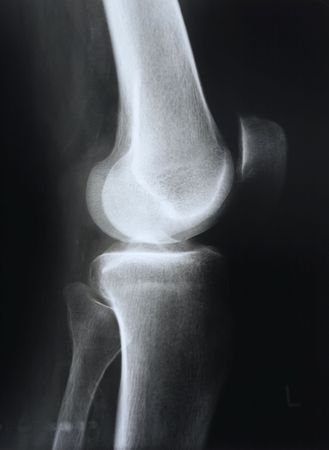
X-ray of knee
favorite
Коллекция по умолчанию
Коллекция по умолчанию
Создать новую
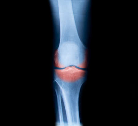
X-ray with osteoarthritis in the knee
favorite
Коллекция по умолчанию
Коллекция по умолчанию
Создать новую
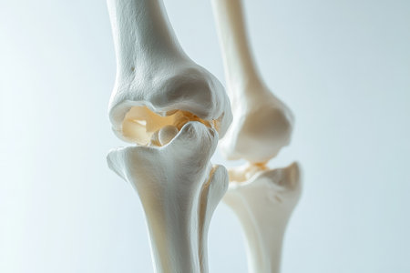
A translucent close-up of the knee joint in a human skeleton model, showing its complex structure in a sleek and minimalistic design, set on a crisp white background.
favorite
Коллекция по умолчанию
Коллекция по умолчанию
Создать новую
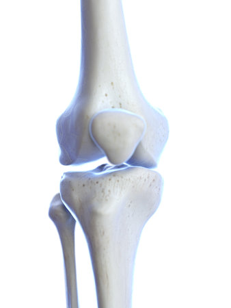
3d rendered medically accurate illustration of the knee joint
favorite
Коллекция по умолчанию
Коллекция по умолчанию
Создать новую
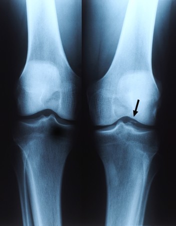
X ray photo of human knee
favorite
Коллекция по умолчанию
Коллекция по умолчанию
Создать новую
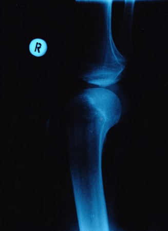
X-ray of the knee-joints over a black background.
favorite
Коллекция по умолчанию
Коллекция по умолчанию
Создать новую

lower leg x-ray of a 48 year old female with a spiral fracture of the distal tibia
favorite
Коллекция по умолчанию
Коллекция по умолчанию
Создать новую

knee with total replacement x-ray image on black background
favorite
Коллекция по умолчанию
Коллекция по умолчанию
Создать новую
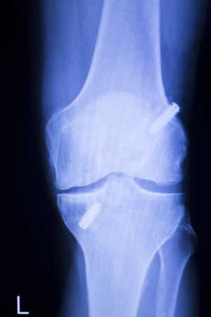
Knee joint implant screw xray showing in medical orthpodedic traumatology scan.
favorite
Коллекция по умолчанию
Коллекция по умолчанию
Создать новую
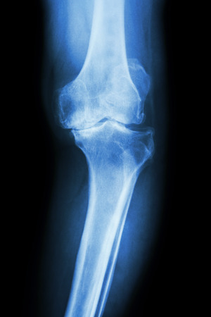
film x-ray knee AP of osteoarthritis knee patient (OA knee)
favorite
Коллекция по умолчанию
Коллекция по умолчанию
Создать новую
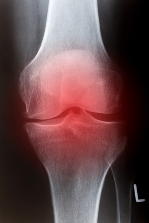
Human knee in front in x-ray
favorite
Коллекция по умолчанию
Коллекция по умолчанию
Создать новую
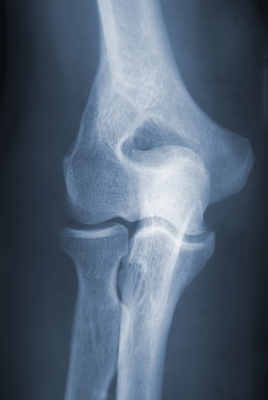
Arm Elbow Joint Vintage 1937 X-Ray
favorite
Коллекция по умолчанию
Коллекция по умолчанию
Создать новую
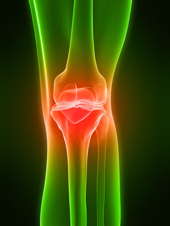
painful knee
favorite
Коллекция по умолчанию
Коллекция по умолчанию
Создать новую

Vector illustration of the human knee joint anatomy
favorite
Коллекция по умолчанию
Коллекция по умолчанию
Создать новую
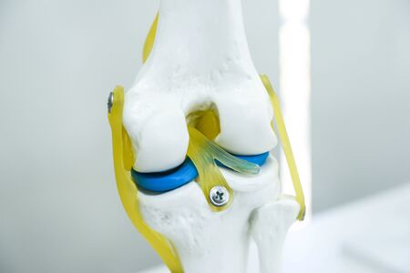
Knee bone model in the hospital for study of knee components
favorite
Коллекция по умолчанию
Коллекция по умолчанию
Создать новую
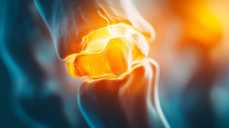
Human Knee Joint Inflammation A Detailed, Close-Up, Anatomical View of Bone Structure.
favorite
Коллекция по умолчанию
Коллекция по умолчанию
Создать новую
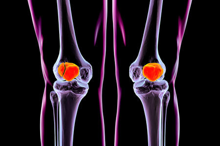
Bipartite patella and normal patella, 3D illustration. The kneecap has an unfused accessory ossification center, and remains as two separate bone fragments instead of fusing completely.
favorite
Коллекция по умолчанию
Коллекция по умолчанию
Создать новую

Closeup back side view of one beautiful sexual young naked woman with flexible straight bare body and buttocks in panties standing in studio on black background, vertical picture
favorite
Коллекция по умолчанию
Коллекция по умолчанию
Создать новую

Human joint prosthesis , metal implant
favorite
Коллекция по умолчанию
Коллекция по умолчанию
Создать новую

Left human knee laterally, x-ray image without any findings
favorite
Коллекция по умолчанию
Коллекция по умолчанию
Создать новую
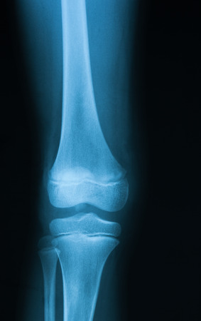
X-ray image of knee joint, AP view.
favorite
Коллекция по умолчанию
Коллекция по умолчанию
Создать новую
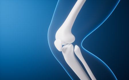
Leg bone in the transparent human body, 3d rendering. 3D illustration.
favorite
Коллекция по умолчанию
Коллекция по умолчанию
Создать новую
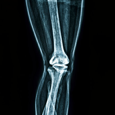
Osteoarthritis Left knee film x-ray AP (anterior - posterior) of knee show narrow joint space
favorite
Коллекция по умолчанию
Коллекция по умолчанию
Создать новую
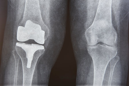
Knee cap replacement xrays. Titanium implant. Osteoarthritis. Articular cartilage
favorite
Коллекция по умолчанию
Коллекция по умолчанию
Создать новую
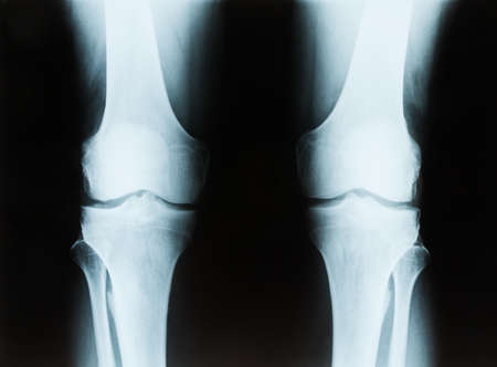
X-ray of a senior male right and left knee showing tibia and fibula bones of both legs
favorite
Коллекция по умолчанию
Коллекция по умолчанию
Создать новую
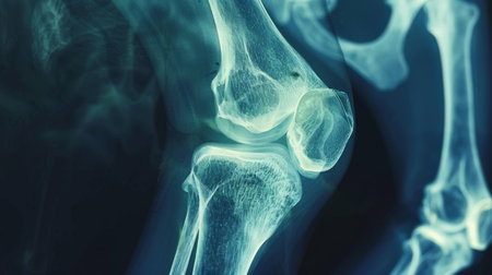
A film x-ray of left knee lateral view shown fracture of knee cap(patella) bone. The plain film of femur on dark background with copy space.Medical concept.Human imaging technology.
favorite
Коллекция по умолчанию
Коллекция по умолчанию
Создать новую
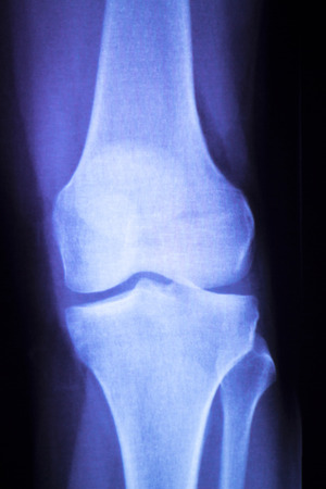
Knee joint meniscus x-ray test scan results photo showing injury and pain for orthopedic surgery and Traumatology surgical treatment.
favorite
Коллекция по умолчанию
Коллекция по умолчанию
Создать новую
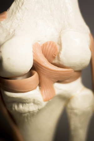
Knee joints meniscus tendons plastic teaching model for taumatology and orthopedics.
favorite
Коллекция по умолчанию
Коллекция по умолчанию
Создать новую
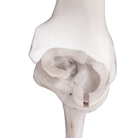
3d rendered medically accurate illustration of a torn medial miniscus
favorite
Коллекция по умолчанию
Коллекция по умолчанию
Создать новую
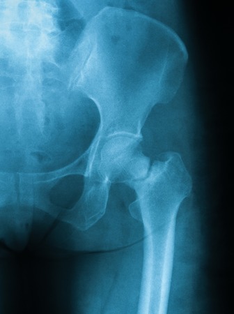
X-ray image of hip joint, AP view. Showing femural neck fracture
favorite
Коллекция по умолчанию
Коллекция по умолчанию
Создать новую
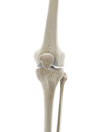
3d rendered medically accurate illustration of the skeletal knee
favorite
Коллекция по умолчанию
Коллекция по умолчанию
Создать новую
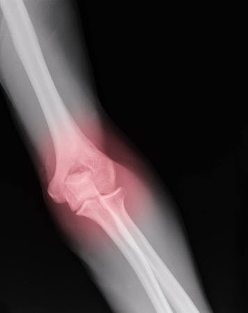
xray of an arm with elbow joint visible and red pain circle
favorite
Коллекция по умолчанию
Коллекция по умолчанию
Создать новую
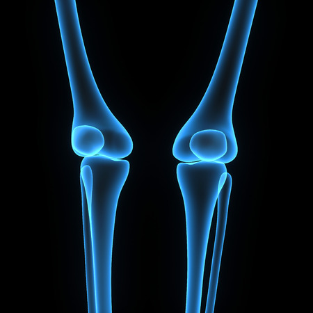
Knee joints
favorite
Коллекция по умолчанию
Коллекция по умолчанию
Создать новую

Inflamed Human Knee Joint A Detailed Anatomical View with Highlighted Cartilage and Bone Structure
favorite
Коллекция по умолчанию
Коллекция по умолчанию
Создать новую
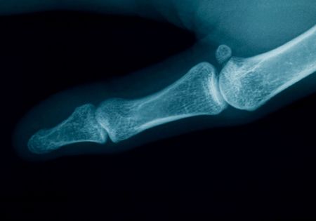
radiography of a middle aged woman finger
favorite
Коллекция по умолчанию
Коллекция по умолчанию
Создать новую

Osteoarthritis Knee ( OA Knee ) ( Film x-ray both knee with arthritis of knee joint : narrow knee joint space ) ( Medical and Science background )
favorite
Коллекция по умолчанию
Коллекция по умолчанию
Создать новую
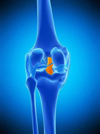
medically accurate illustration of the posterior cruciate ligament
favorite
Коллекция по умолчанию
Коллекция по умолчанию
Создать новую
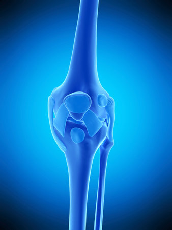
medically accurate illustration of the knee ligaments
favorite
Коллекция по умолчанию
Коллекция по умолчанию
Создать новую

X-ray orthopedic medical CAT scan of painful knee meniscus injury leg in traumatology hospital clinic.
favorite
Коллекция по умолчанию
Коллекция по умолчанию
Создать новую

medically accurate illustration of the fibular collateral ligament
favorite
Коллекция по умолчанию
Коллекция по умолчанию
Создать новую
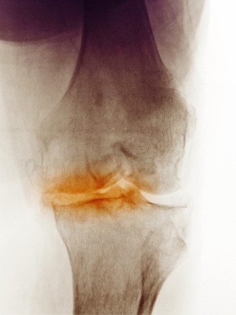
X-ray of the knee of a 83 year old woman showing degenerative arthritis. This woman was scheduled for a knee replacement.
favorite
Коллекция по умолчанию
Коллекция по умолчанию
Создать новую

collection knee x-ray image
favorite
Коллекция по умолчанию
Коллекция по умолчанию
Создать новую
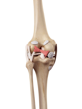
medical accurate illustration of the oblique politeal ligament
favorite
Коллекция по умолчанию
Коллекция по умолчанию
Создать новую

Ankle feet & knee joint X-ray human photo film
favorite
Коллекция по умолчанию
Коллекция по умолчанию
Создать новую
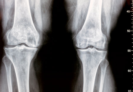
Plain X ray of both knee joints shows apparent joint osteoarthritis according to Kellgren and Lawrence system for classification of osteoarthritis with definite osteophytes and joint space narrowing
favorite
Коллекция по умолчанию
Коллекция по умолчанию
Создать новую
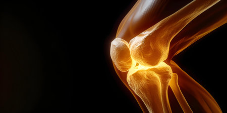
Detailed D illustration of a knee in pain showing medical accuracy. Concept Knee Pain Anatomy, Detailed Illustration, Medical Accuracy, 3D Rendering
favorite
Коллекция по умолчанию
Коллекция по умолчанию
Создать новую

Human Knee joint pain, medical concept. 3D Rendering
favorite
Коллекция по умолчанию
Коллекция по умолчанию
Создать новую
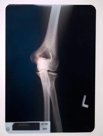
X-ray of a knee
favorite
Коллекция по умолчанию
Коллекция по умолчанию
Создать новую
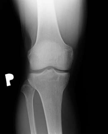
Preventive and control medical examination. X-ray of the right man's knee. Visible early degenerative changes. Front view.
favorite
Коллекция по умолчанию
Коллекция по умолчанию
Создать новую
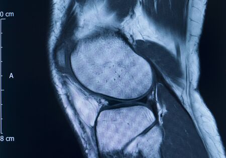
Knee sports injury mri mcl grade 2 tear magnetic resonance imaging orthopedic traumatology scan.
favorite
Коллекция по умолчанию
Коллекция по умолчанию
Создать новую
abstract computer graphic background art wallpaper
favorite
Коллекция по умолчанию
Коллекция по умолчанию
Создать новую

medically accurate 3d illustration of the human knee
favorite
Коллекция по умолчанию
Коллекция по умолчанию
Создать новую
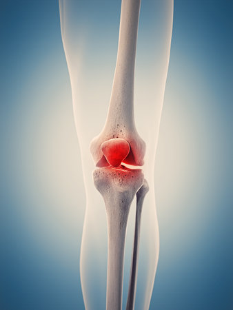
medically accurate 3d illustration of the painful knee
favorite
Коллекция по умолчанию
Коллекция по умолчанию
Создать новую
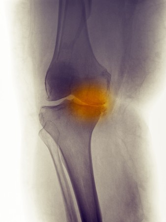
Knee x-ray of a 60 year old woman showing degenerative joint disease with severe narrowing of the medial joint line of the knee
favorite
Коллекция по умолчанию
Коллекция по умолчанию
Создать новую

film x-ray both knee joints and pain area.
favorite
Коллекция по умолчанию
Коллекция по умолчанию
Создать новую
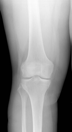
Image Of Knee X-Ray. Detecting Radiographic Knee Osteoarthritis.
favorite
Коллекция по умолчанию
Коллекция по умолчанию
Создать новую
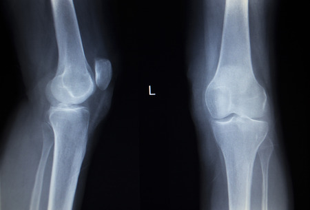
X-ray orthopedic medical CAT scan of painful knee meniscus injury leg in traumatology hospital clinic.
favorite
Коллекция по умолчанию
Коллекция по умолчанию
Создать новую

man's leg
favorite
Коллекция по умолчанию
Коллекция по умолчанию
Создать новую
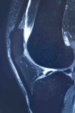
Knee sports injury mri mcl grade 2 tear magnetic resonance imaging orthopedic traumatology scan.
favorite
Коллекция по умолчанию
Коллекция по умолчанию
Создать новую
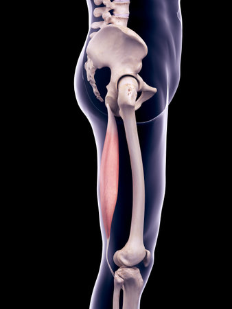
medically accurate illustration of the semitendinosus
favorite
Коллекция по умолчанию
Коллекция по умолчанию
Создать новую
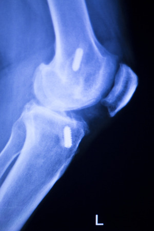
Knee joint implant screw xray showing in medical orthpodedic traumatology scan.
favorite
Коллекция по умолчанию
Коллекция по умолчанию
Создать новую
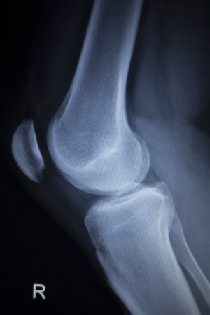
X-ray orthopedic medical CAT scan of painful knee meniscus injury leg in traumatology hospital clinic.
favorite
Коллекция по умолчанию
Коллекция по умолчанию
Создать новую
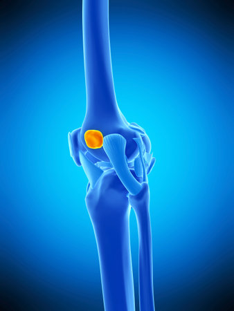
medically accurate illustration of the iliotibial tract bursa
favorite
Коллекция по умолчанию
Коллекция по умолчанию
Создать новую

CT knee joint Coronal view isolated on black background showing fracture Femur bone.
favorite
Коллекция по умолчанию
Коллекция по умолчанию
Создать новую
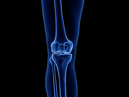
3d rendered medically accurate illustration of the healthy knee joint
favorite
Коллекция по умолчанию
Коллекция по умолчанию
Создать новую

Close up augmented reality animation showing joint and knee pain from leg trauma or arthritis
favorite
Коллекция по умолчанию
Коллекция по умолчанию
Создать новую

Human Knee Joint Inflammation Anatomical Close-Up, Highlighted Pain, Blue Background
favorite
Коллекция по умолчанию
Коллекция по умолчанию
Создать новую
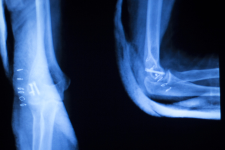
Othopedics and Traumatology surgical implant arm and elbow xray test scan results showing titanium metal plate and screws.
favorite
Коллекция по умолчанию
Коллекция по умолчанию
Создать новую

x-ray of a human knee with prothesis
favorite
Коллекция по умолчанию
Коллекция по умолчанию
Создать новую
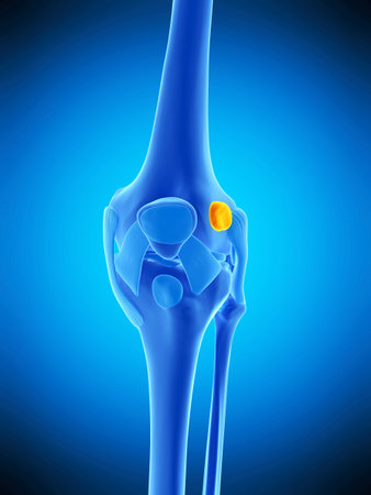
medically accurate illustration of the iliotibial tract bursa
favorite
Коллекция по умолчанию
Коллекция по умолчанию
Создать новую
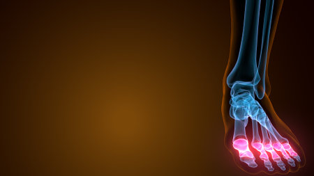
X-ray of human foot on a dark background with copy space
favorite
Коллекция по умолчанию
Коллекция по умолчанию
Создать новую
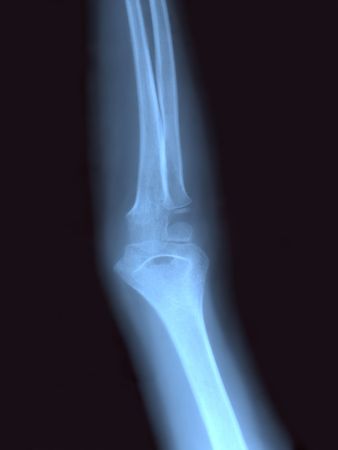
Arm X RAY
favorite
Коллекция по умолчанию
Коллекция по умолчанию
Создать новую
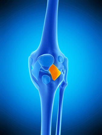
medically accurate illustration of the lateral patellar ligament
favorite
Коллекция по умолчанию
Коллекция по умолчанию
Создать новую
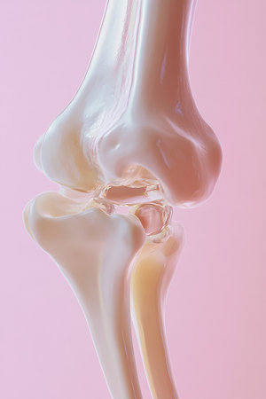
A high-resolution close-up of a translucent 3D model of a human skeleton's knee joint, illustrating its anatomy with a minimalistic approach, complemented by a pastel-colored background.
favorite
Коллекция по умолчанию
Коллекция по умолчанию
Создать новую
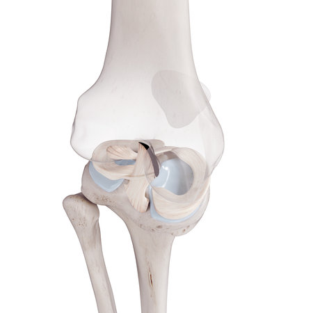
3d rendered medically accurate illustration of the cruciate ligaments and menisci
favorite
Коллекция по умолчанию
Коллекция по умолчанию
Создать новую

flat icon with long shadow human bone
favorite
Коллекция по умолчанию
Коллекция по умолчанию
Создать новую
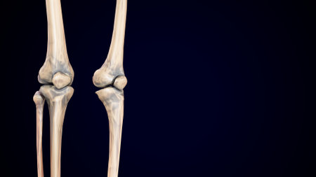
X-ray of human knee joint on blue background. Medical and healthcare concept.
favorite
Коллекция по умолчанию
Коллекция по умолчанию
Создать новую

X-ray picture showing knee joints
favorite
Коллекция по умолчанию
Коллекция по умолчанию
Создать новую

Human skeleton anatomy articularis Bone 3D Rendering For Medical Concept
favorite
Коллекция по умолчанию
Коллекция по умолчанию
Создать новую

Augmented reality animation showing person with joint and knee pain from leg trauma or arthritis
favorite
Коллекция по умолчанию
Коллекция по умолчанию
Создать новую
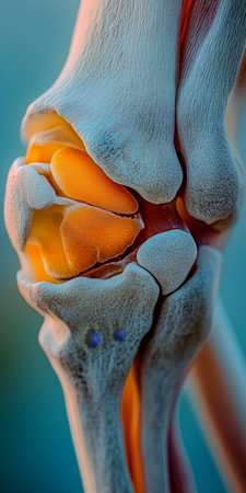
Close-Up View of an Inflamed Human Knee Joint, Highlighting Cartilage and Bone Structure
favorite
Коллекция по умолчанию
Коллекция по умолчанию
Создать новую
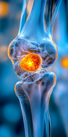
Inflamed Human Knee Joint, Anatomical Illustration, Vibrant Color, Gradient Blue Background
favorite
Коллекция по умолчанию
Коллекция по умолчанию
Создать новую
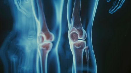
Film x-ray human's knee joints
favorite
Коллекция по умолчанию
Коллекция по умолчанию
Создать новую
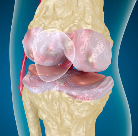
Osteoarthritis Knee
favorite
Коллекция по умолчанию
Коллекция по умолчанию
Создать новую
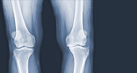
Film x-ray of human knee Osteoarthritis of the Knee normal ligaments Medical image concept.
favorite
Коллекция по умолчанию
Коллекция по умолчанию
Создать новую

CT scan of elbow joint 3d rendering .
favorite
Коллекция по умолчанию
Коллекция по умолчанию
Создать новую
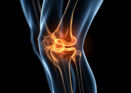
Osteoarthritis of the knee and hand. Healthcare concept image
favorite
Коллекция по умолчанию
Коллекция по умолчанию
Создать новую
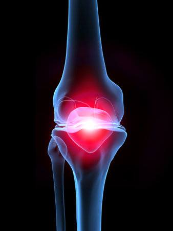
skeletal knee with pain
favorite
Коллекция по умолчанию
Коллекция по умолчанию
Создать новую
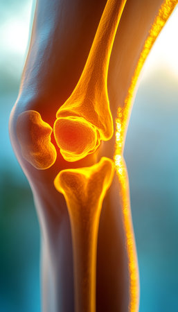
Inflamed Human Knee Joint A 3D Rendering of Anatomical Detail on Blue Gradient Background
favorite
Коллекция по умолчанию
Коллекция по умолчанию
Создать новую

Scanogram is a Full-length standing AP radiograph of both lower extremities including the hip, knee, and ankle.
favorite
Коллекция по умолчанию
Коллекция по умолчанию
Создать новую
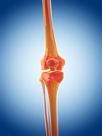
medically accurate illustration of the knee joint
favorite
Коллекция по умолчанию
Коллекция по умолчанию
Создать новую

medically accurate illustration of the knee anatomy
favorite
Коллекция по умолчанию
Коллекция по умолчанию
Создать новую

X-ray of both human knee
favorite
Коллекция по умолчанию
Коллекция по умолчанию
Создать новую

X-ray orthopedic medical CAT scan of painful knee meniscus injury leg in Traumatology hospital clinic with prosthetics Trauma implant.
favorite
Коллекция по умолчанию
Коллекция по умолчанию
Создать новую
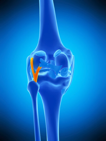
medically accurate illustration of the arcuate politeal ligament
favorite
Коллекция по умолчанию
Коллекция по умолчанию
Создать новую
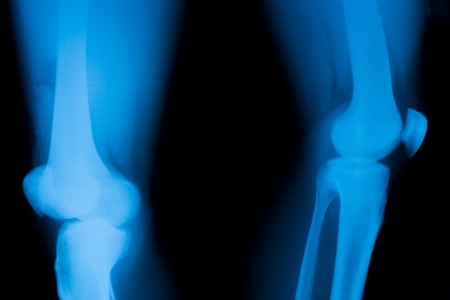
Patella dislocation.
favorite
Коллекция по умолчанию
Коллекция по умолчанию
Создать новую
Поддержка
Copyright © Legion-Media.


